Interstitial Lung Disease Radiographics
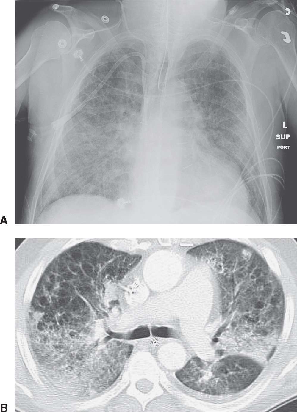
Interstitial lung disease radiographics. Prominent interstial markings on a regular chest x-ray are often due to x-ray technique. Interstitial Lung Disease Etiology Known Etiology Unknown aka idiopathic Unclassifiable Autoimmune disease - RA SSc Sjogrens IIM Environmental ILD - Hypersensitivity pneumonitis Occupational ILD - AsbestosisSilicosis Drug-induced ILD - AmioMTXChemo Smoking-related - Desquamative interstitial pneumonia - Respiratory bronchiolitis-ILD Chronic Fibrosing. Interstitial lung disease ILD in pediatric patients is different from that in adults with a vast array of pathologic conditions unique to childhood varied modes of presentation and a different range of radiologic appearances.
Lymphoid interstitial pneumonia LIP. The clinical assessment of patients with suspected ILD includes a thorough history physical examination and pulmonary function testing. Respiratory bronchiolitisassociated interstitial lung disease RB-ILD.
Although rare childhood ILD chILD is associated with significant morbidity and mortality most notably in conditions. Send thanks to the doctor. IIPs include seven entities.
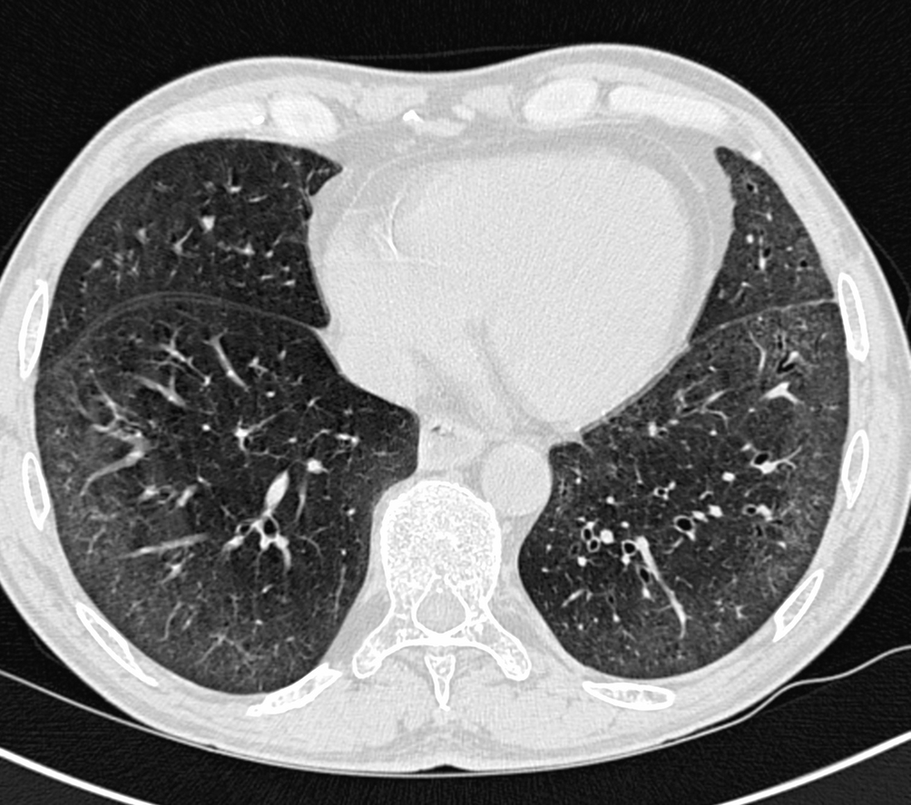
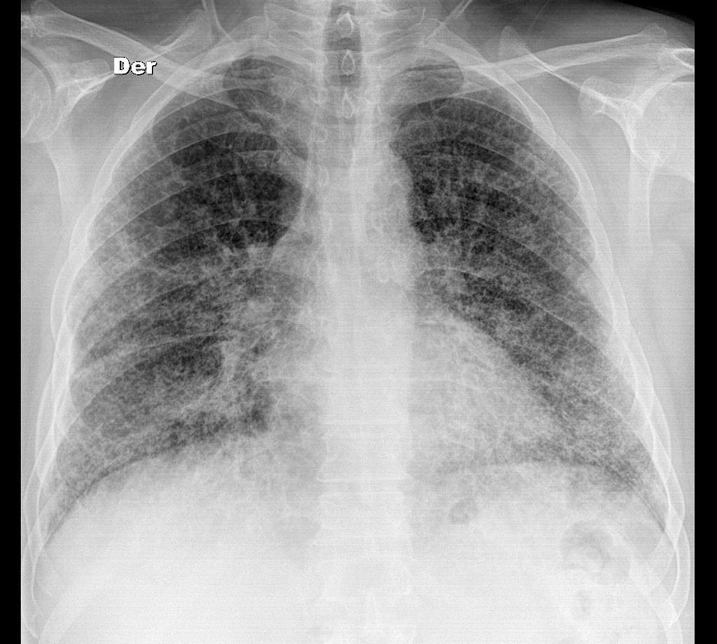
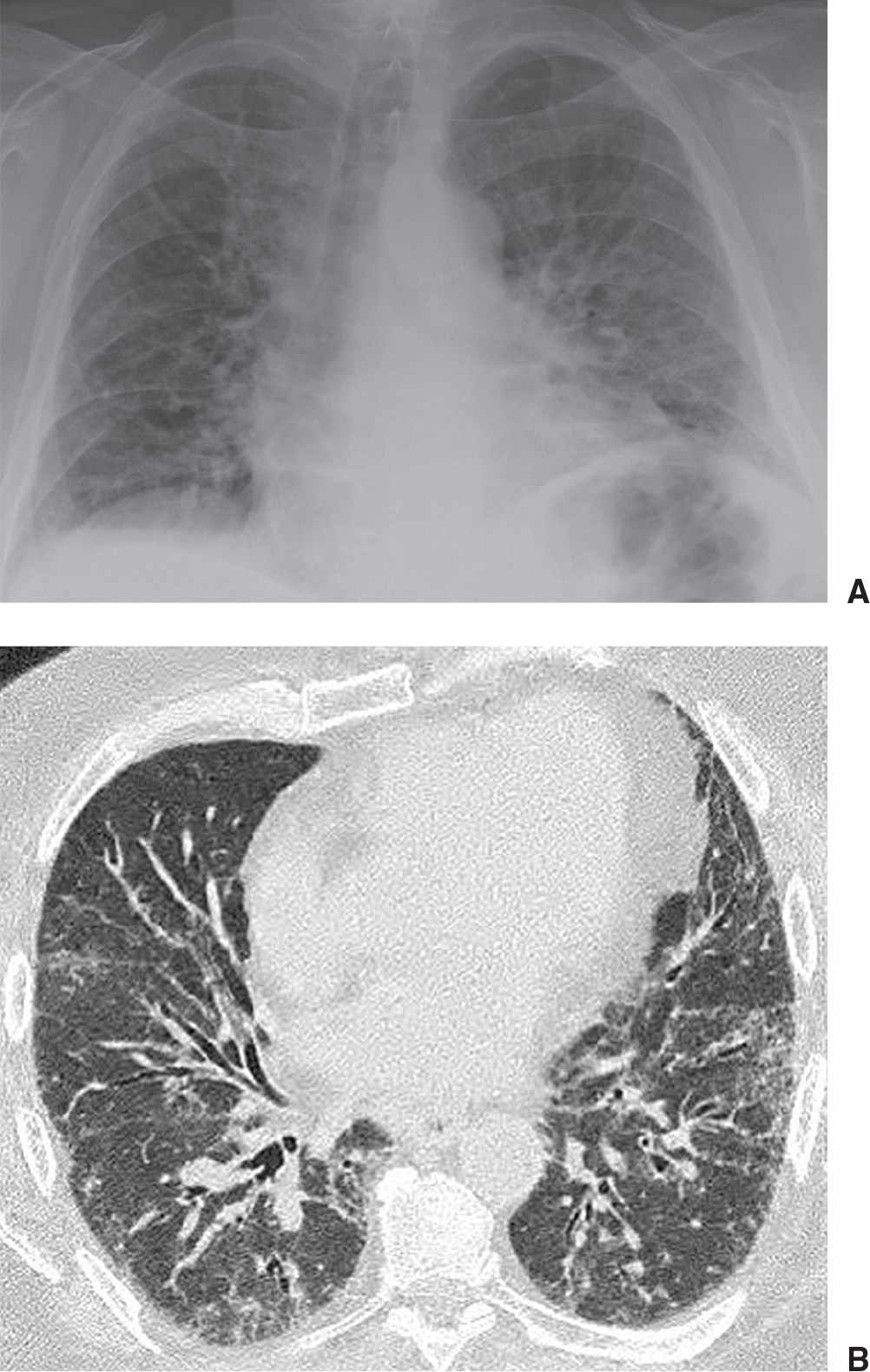
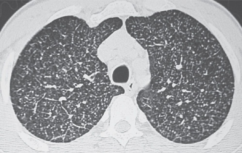
Reticular nodular high and low attenuation table. An interstitial lung pattern is a regular descriptive term used when reporting a plain chest radiograph. The interstitial pneumonias are a group of heterogeneous nonneoplastic lung diseases that may be idiopathic or associated with an underlying abnormality.
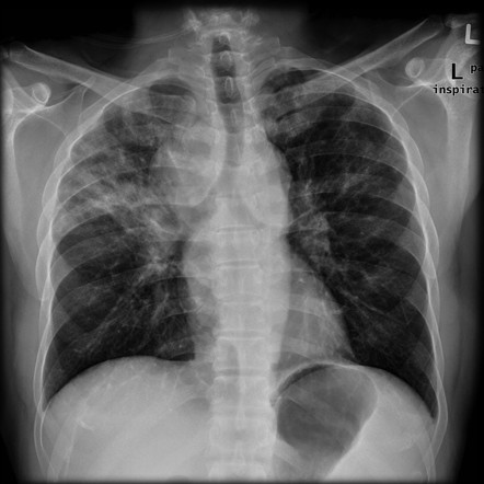
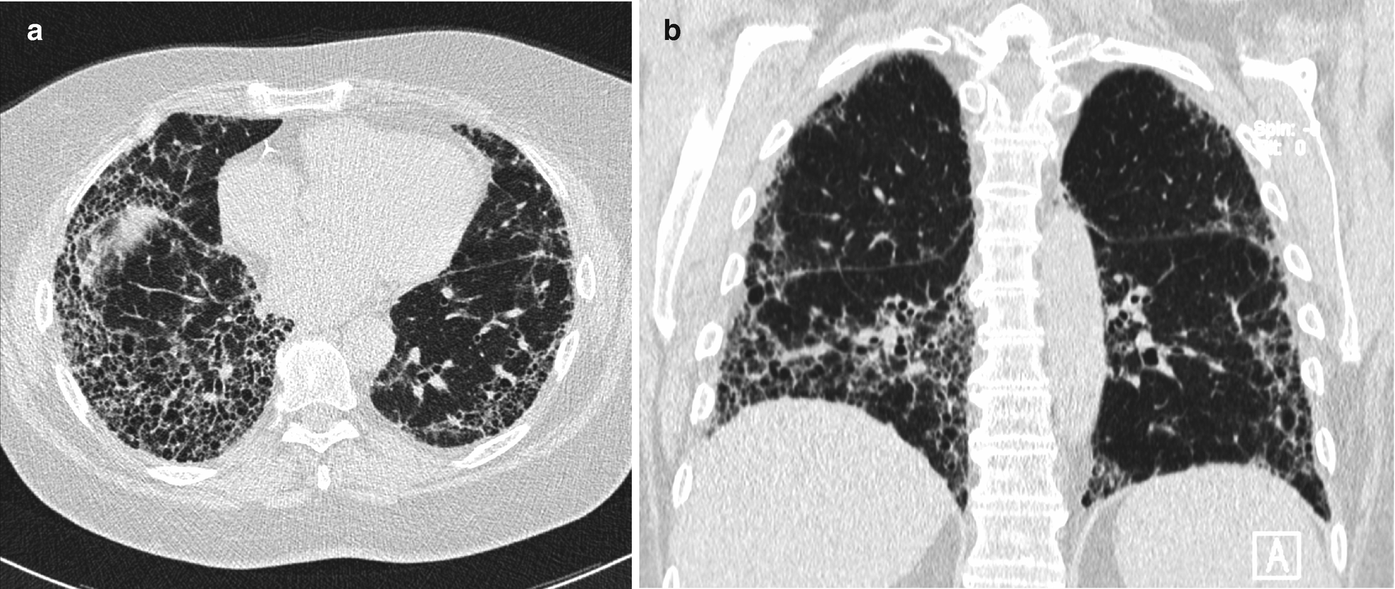
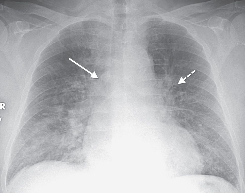
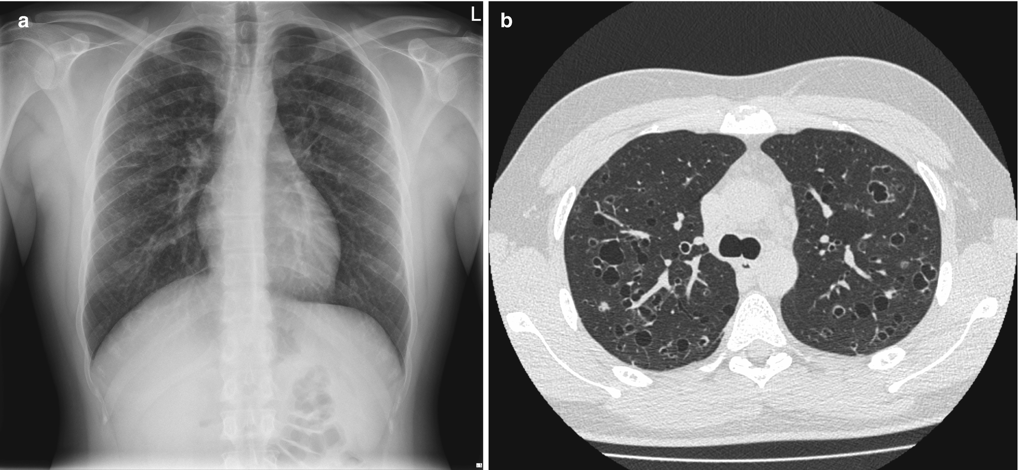
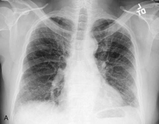
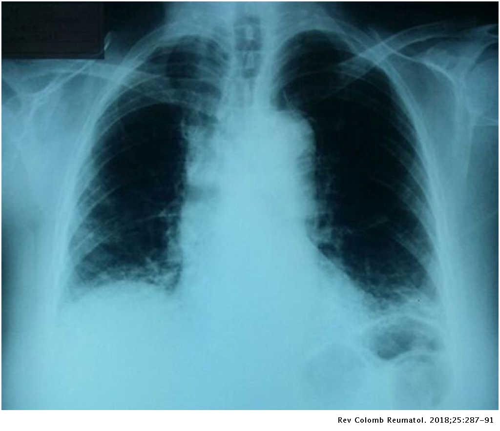
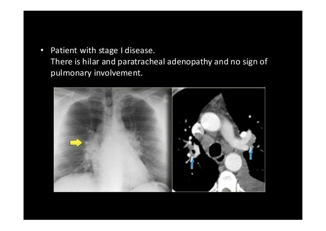
On a Chest X-Ray it can be very difficult to determine whether there is interstitial lung disease and what kind of pattern we are dealing with. A graphic or morphometric classification is a better approach and is enumerated in Box 7-3. Although they share some features in common they also exhibit diverse pulmonary manifestations.
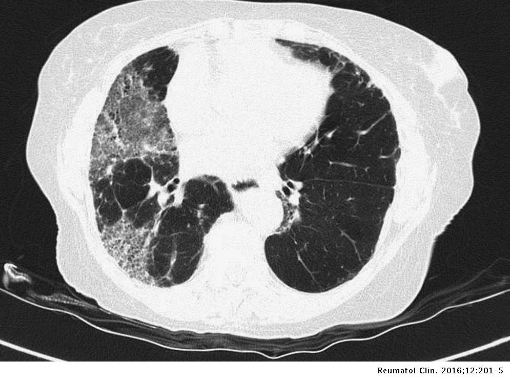
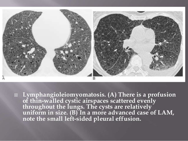
Radiologic and histopathologic findings. The interstitial pneumonias are a heterogeneous group of diffuse parenchymal lung diseases with diverse imaging manifestations clinical features and outcomes. Knowledge of both the radiological and clinical appearance of these more common interstitial lung diseases is therefore important for recognizing them in the daily practice and including them in the differential diagnosis.
It is the result of the age-old attempt to make the distinction between an interstitial and airspace alveolar process to narrow the differential diagnosis. To diagnose interstial lung disease you should have a cat sc.
The clinical assessment of patients with suspected ILD includes a thorough history physical examination and pulmonary function testing.
Cryptogenic organizing pneumonia COP. They may be idiopathic or secondary to some other cause. IPF affects around 3 million people worldwide with incidence increasing dramatically with age. Knowledge of both the radiological and clinical appearance of these more common interstitial lung diseases is therefore important for recognizing them in the daily practice and including them in the differential diagnosis. Nonspecific interstitial pneumonia NSIP. To diagnose interstial lung disease you should have a cat sc. Interstitial lung disease ILD in RA RA-ILD and other types of connective tissue diseases CTDs is categorized using the international consensus. Although they share some features in common they also exhibit diverse pulmonary manifestations. IPF is characterized by progressive worsening of dyspnea and lung.
Interstitial lung diseases associated with collagen vascular diseases. A graphic or morphometric classification is a better approach and is enumerated in Box 7-3. View 1 more answer. Desquamative interstitial pneumonia DIP. Reticular nodular high and low attenuation table. Prominent interstial markings on a regular chest x-ray are often due to x-ray technique. Lymphoid interstitial pneumonia LIP.
















Post a Comment for "Interstitial Lung Disease Radiographics"